Precision experts rely on.
 Advice wanted

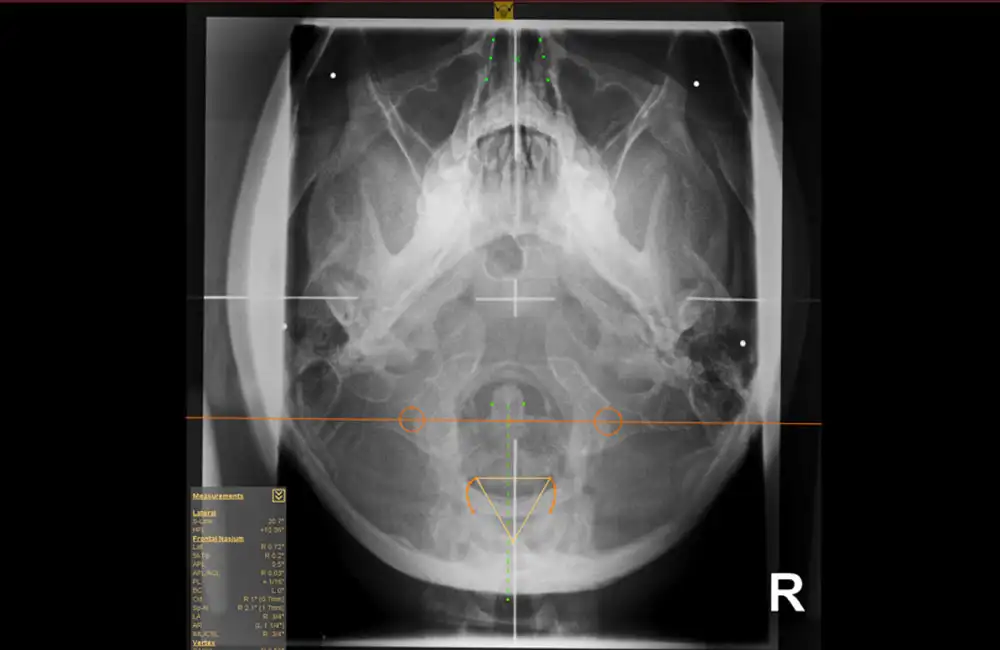
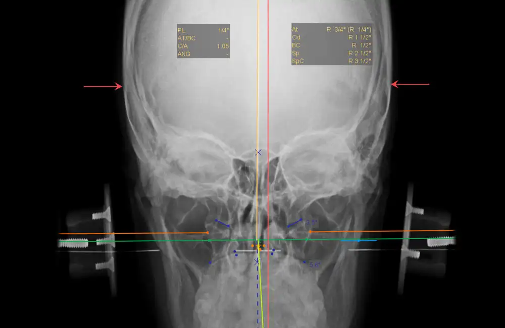
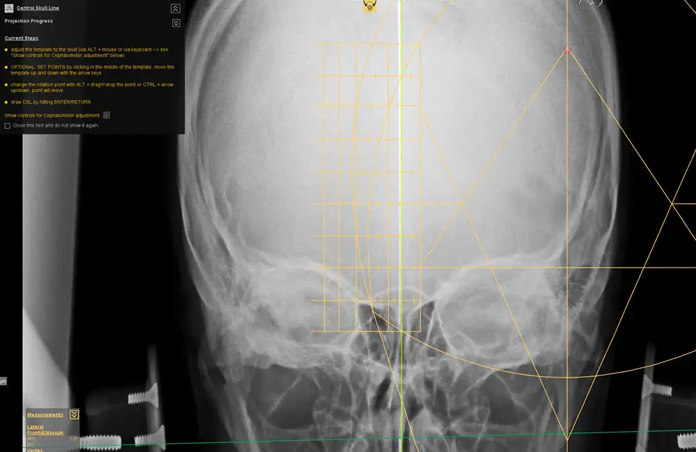
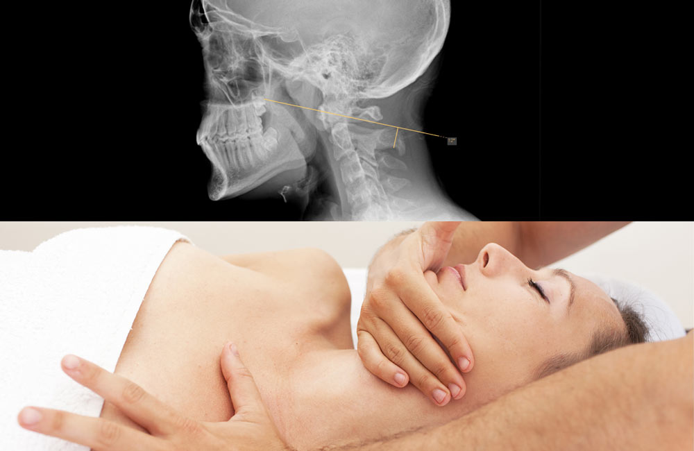
The dicomPACS® Upper Cervical (NUCCA) Chiropractic Tool Set was developed in close collaboration with leading NUCCA experts from the United States and Canada. It provides everything required for fast and highly precise diagnostics in the upper cervical spine. Specialized NUCCA templates such as the cephalometer, grid, circumscale, and relatoscope support a familiar workflow while ensuring maximum accuracy.
Using manual point marking, the software automatically generates midlines, reference points, curves, and angles, significantly improving the efficiency of diagnosis and treatment planning. The NUCCA Tool Set is complemented by all essential standard chiropractic tools, including distance and angle measurements, Cobb angles, and marker points—delivering a comprehensive, professional NUCCA analysis within a single, integrated solution.
Cephalometer, grid, circumscale, and relatoscope for precise analysis with a familiar workflow.
Automatic generation of midlines, reference points, curves, and angles based on manual point marking.
NUCCA-specific lines and structures such as the atlas plane line and S-line for accurate diagnostics.
All essential measurement and marking functions, including angle, distance, and Cobb measurements.
Our support team is at your side – by telephone and remote maintenance. We ensure the smooth operation of your software.
Overview of services
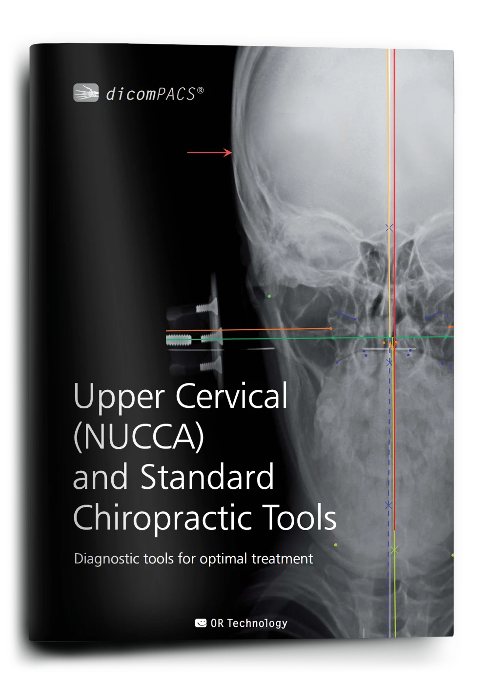
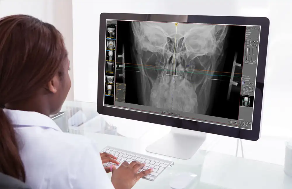
dicomPACS® Upper Cervical (NUCCA) and Standard Chiropractic Tools
Diagnostic tools for optimal chiropractic reporting
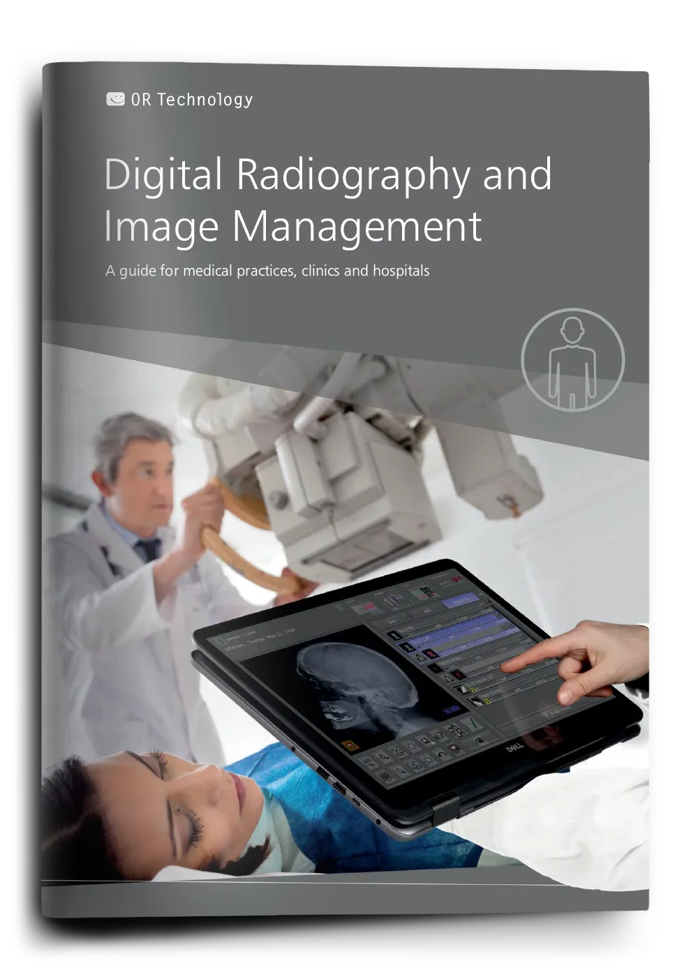
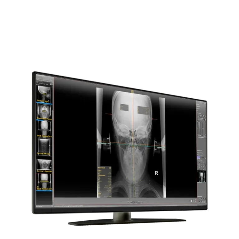
Digital X-ray and image management
The guide for medical practices and clinics
dicomPACS®MobileView is a web-based system that enables location-independent display and editing of radiological image data on mobile devices.
Learn more (PDF)The web-based viewer contains all the necessary basic functions for viewing and editing images, which can be done on mobile devices regardless of the browser used.
Learn moreThe prosthesis documentation module enables preoperative planning and documentation. Digital templates can be superimposed on the X-ray image or foil prosthesis templates can be used.
Find out more (contact form)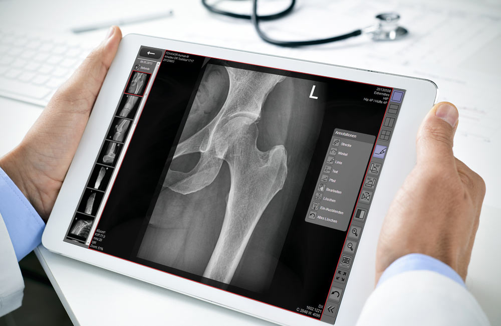
dicomPACS®MobileView is a web-based system that enables location-independent display and editing of radiological image data on mobile devices.
Learn more (PDF)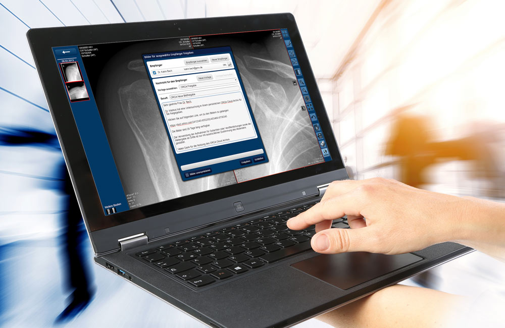
The web-based viewer contains all the necessary basic functions for viewing and editing images, which can be done on mobile devices regardless of the browser used.
Learn more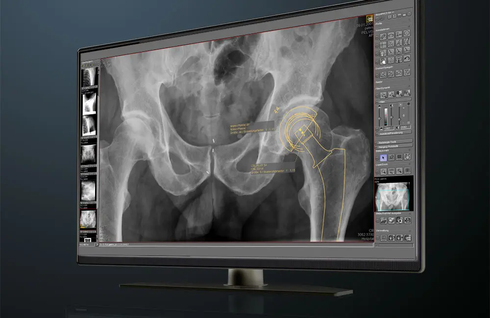
The prosthesis documentation module enables preoperative planning and documentation. Digital templates can be superimposed on the X-ray image or foil prosthesis templates can be used.
Find out more (contact form)
The Vital PostureTM Clinic
Doctor Jeff Scholten is a great advocate of the NUCCA software development and works with dicomPACS® Upper Cervical Chiropractic Tools. What also impressed him was that could be updated to a fulldicomPACS® analysis tool for ALL diagnostic imaging needs even MRl or CT images…) “The software’s modular system is so much more than just another analysis tool. It supports my workflow and makes it easier.”Reference (PDF)
Subject to technical changes. All details regarding disclaimer, copyrights and our legal requirements can be found at here.
OR Technology (Oehm und Rehbein GmbH)
Neptunallee 7c
18057 Rostock,
Germany
Celtic SMR ltd.
Frederick House, Hayston View,
Johnston SA62 3AQ,
United Kingdom
Celtic SMR ltd.
Sovereign Court, Wyrefields,
Poulton Industrial Estate,
Poulton-le-Fylde FY6 8JX,
United Kingdom
Vetequip
Unit C1, Clane Business Park,
College Road, Clane,
Co. Kildare, W91 VK84
Ireland
© 2026 OR Technology | All rights reserved.