


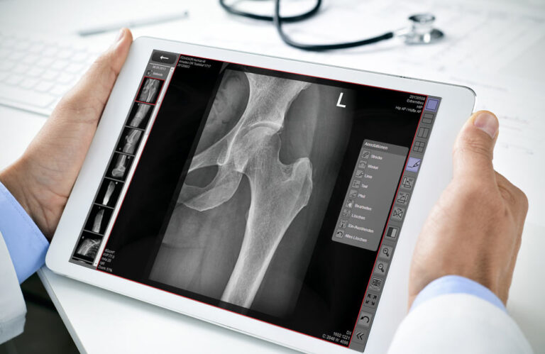
Mobile image viewing
dicomPACS®MobileView
dicomPACS® MobileView is a web-based system that enables location-independent viewing and editing of radiological image data on mobile devices.


The heart of your X-ray system: Professional acquisition software for your X-ray images
 Advice wanted

ORmed™ delivers professional X-ray images in outstanding quality. The software controls your X-ray system, optimises images organ-specifically, and enables efficient workflows. With intuitive operation, a multimedia X-ray assistant, and easy integration into management systems, ORmed™ ensures maximum precision – suitable for small clinics and large hospitals.
User-friendly, fast, and efficient – modern graphical interface, touchscreen operation, efficient workflows, 400 preconfigured projections
Easy connectivity – automatic patient data transfer, DICOM Worklist, REST API, HIS/RIS integration
Customizable, flexible, and efficient – free configuration of body parts, macros for examinations, adding images afterwards
Safe and reliable – multimedia X-ray assistant, fast registration of emergency patients, switch between examinations, support & remote maintenance
Our support team is at your side – by telephone and remote maintenance. We ensure the smooth operation of your software.
Overview of services

Acquisition, reporting and archiving of image sequences
(Available as an extension.)
Dynamic X-ray imaging in ORmed™ enables continuous visualisation of the region being examined on the diagnostic display using pulsed X-rays.
This method is particularly suited to visualising movement, such as respiratory motion and movement of the diaphragm. Functional processes in the thorax can therefore be visualised through short image sequences.
ORmed™ supports dynamic X-ray imaging with high-resolution detectors in a range of sizes.
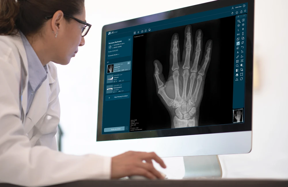
From examination planning to the finished image
ORmed™ supports the entire digital X-ray process: from importing patient data through examination planning and image acquisition to processing and transfer to the PACS.
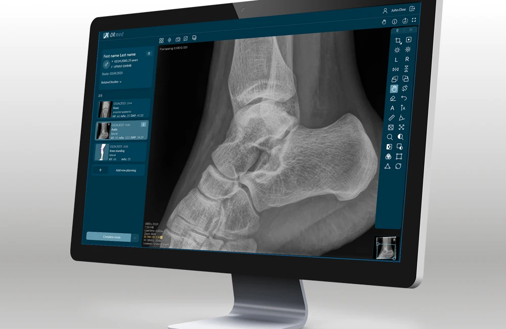
Automatically optimised X-ray images
Cognition Optimised Processing (COP) automatically analyses the acquired image data and adapts processing to the anatomical region and acquisition conditions. High-quality images are therefore generally available immediately after acquisition, without extensive manual post-processing.
Automatic functions include:
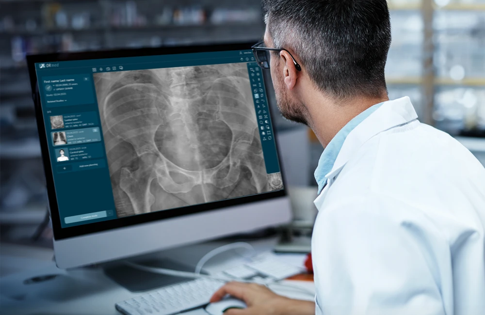
A comprehensive range of integrated tools
Acquired images can be reviewed and edited directly in ORmed™. Functions such as zoom, window and level adjustment, length and angle measurements, text and arrow annotations, rotation, mirroring and the display of multiple images support image review and editing during the examination workflow.
Completed examinations can be automatically transferred to the PACS or exported as image files or DICOM files.
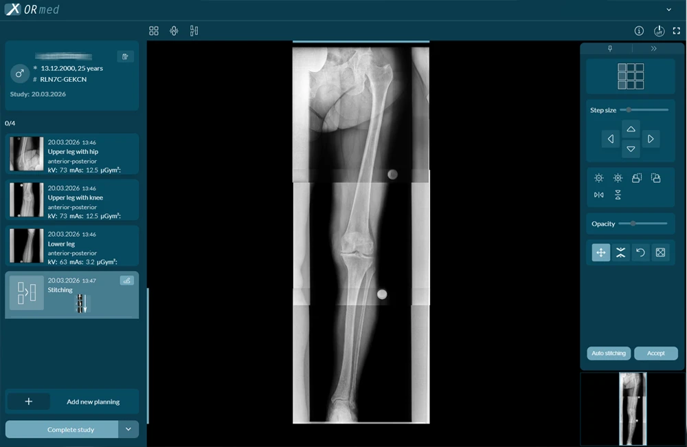
ORmed™ automatically combines multiple overlapping X-ray images into an anatomically consistent composite image. This provides a continuous view of anatomical regions that extend beyond the size of the detector.
Macros simplify the planning of long-leg or spinal X-rays. Automatic image alignment supports stitching, while manual correction options allow individual images to be adjusted precisely.
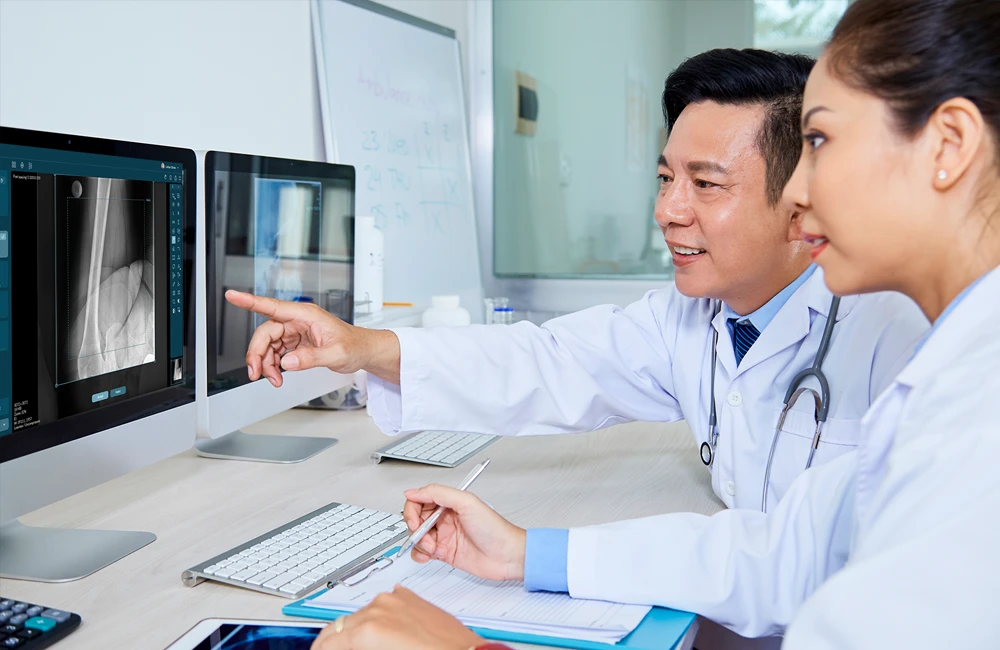
The right addition to your everyday workflow.
Mobile image viewing
dicomPACS® MobileView is a web-based system that enables location-independent viewing and editing of radiological image data on mobile devices.
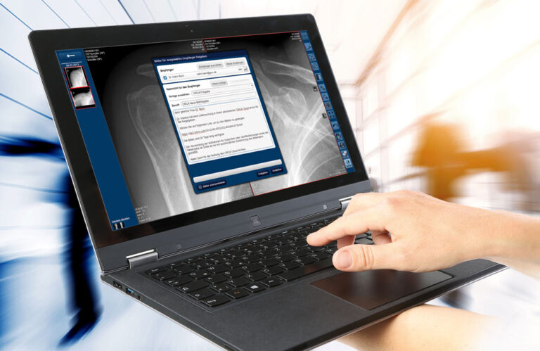
Cloud archiving & sharing
The web-based viewer includes all essential basic functions for viewing and editing images, which can be performed almost browser-independently on mobile devices.
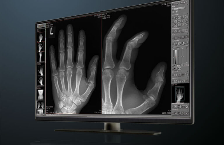
Digital image management
dicomPACS® is a modern and advanced high-tech solution for intelligent image management. All images from X-ray, CT, MRI, and ultrasound, as well as documents such as medical reports, findings, and faxes, can be stored in the digital patient file and retrieved at the touch of a button.
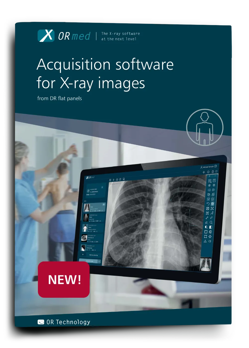
ORmed™ X-ray software on the next level
versatile software solution for X-ray image acquisition
Subject to technical changes. All details regarding disclaimer, copyrights and our legal requirements can be found at here.