![]()
![]()

")


The use of chemicals in traditional analog techniques is associated with occupational health hazards and unpleasant odours. The costs of conventional x-ray archives are high in terms of space, time and money. Digital radiography represents an environmentally friendly and high-quality alternative. Reduced radiation exposure for patients is a further advantage over conventional radiography. Digital and analog imaging systems differ with regard to image production and image display. However, digital X-ray images are subject to the same rules of physics as conventional images. Instead of viewing photographic film using a lightbox, digital images are visualized with a PC and display monitor. Thus, the transition to digital radiography involves only minor changes in established workflows. Patient handling is not affected.
Not interested in a new digital X-ray machine at the moment? No problem!

Your conventional X-ray unit can easily be transitioned to an digital X-ray machine by the addition of a flat panel detector (DR) or a CR scanner in combination with a medical display monitor. While continuing to use your existing radiography system, you can reap the benefits of high-quality digital X-ray images.
 |
We offer you an extensive selection of Digital X-ray machines, mobile X-ray units, protable X-ray suitcases and DR upgrade sets (for your conventional X-ray machine) for orthopaedic and surgical practices, radiology and hospitals as well as medical services including X-ray software (acquisition and diagnostic software from OR Technology) |
Digital direct radiography (DR) produces images of outstanding quality. A flat panel detector directly converts the pattern of incident X-ray energy into electrical signals. Imaging plates, cassettes and X-ray scanners are no longer needed . The X-ray images are available for diagnosis immediately after exposure.

DR systems can build upon existing conventional X-ray equipment. Digital upgrades do not necessarily involve modification of the X-ray machine or X-ray unit. The system‘s Auto Exposure Detection (AED) works without changes to the running system or cable connections. The new 35 x 43 cm X-ray detectors are designed to fit perfectly into the Bucky tray of existing X-ray units.
Direct radiography systems have high throughput and are ideal for emergency medicine and intensive care.
Choose the digital X-ray machine (DR) that fits your needs:
 |
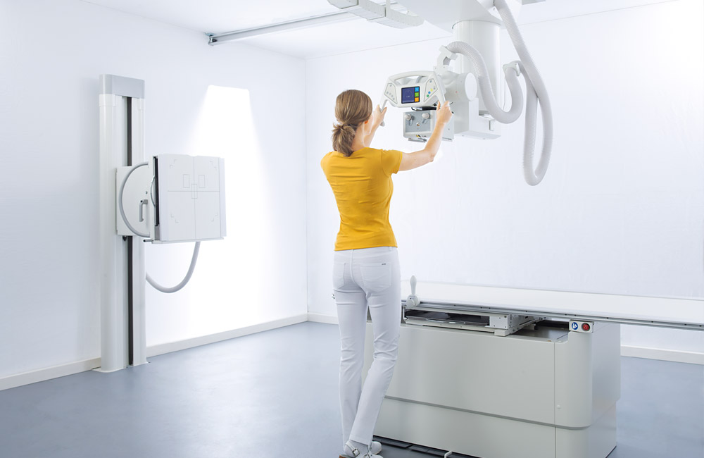
Motorised, digital U-arm X-ray machine Amadeo Z motorised for all X-ray exposures in sitting, standing and lying positionThe compact, digital U-arm X-ray machine is motorised and designed for low ceiling heights from 2.40 m. Five electric motors allow effortless and accurate positioning of the stand and ensure a wide range of exposures. All radiological settings can be utilised, as well as automated whole spine and whole leg exposures (stitching). All important settings and operating operations are carried out via an integrated 10" touchscreen display console. Frequently used unit positions can be predefined on the 60 available programme slots to ensure fast positioning on the patient. |
 |
Amadeo R motorised: Motorised, universal X-ray machine with bucky table and wall stand |
 |
Motorised digital U-arm X-ray machine for all applications in diagnostic radiographyThe Amadeo S motorised is a motorised U-arm X-ray machine for the production of all diagnostic projection radiography images. Both its compact design and the minimum ceiling height of only 2.40 m make the system perfectly suited for small rooms. It includes all necessary components and functions for digital X-ray without cassettes: motorised U-arm system, generator, X-ray detector, PC and the dicomPACS®DX-R acquisition and diagnostic software. |
 |
Amadeo M mini Systems: A lightweight and mobile X-ray machine for wireless, digital X-ray imaging in ambulatory and inpatient care settingsThis mobile, digital X-ray machine is indispensable for in situ radiological examinations when patients cannot be transferred to a hospital. The mobile full X-ray system was developed for doctors and first aid personnel in remote and inaccessible locations, medical aid organisations, as well as ships and offshore platforms. |
 |
Medici DR Systems: Digital retrofits for stationary X-ray machineAre you interested in switching to digital X-ray imaging? If yes, then our Medici system is your best bet! Medici DR systems are available for nearly every X-ray unit manufactured. Choose among a large selection of detector models and sizes to optimally configure your digital X-ray equipment. |
 |
Medici DR Systems: Digital retrofits for mobile X-ray unitsAre you interested upgrading your trusted mobile X-ray machine to produce professional and reproducible digital images? Are you looking to connect your existing X-ray unit to a digital X-ray sensor that is easy to install and straightforward to operate? Choose among the large selection of wireless detector models and sizes in the Medici system to customize your X-ray system. The dicomPACS®DX-R image acquisition software produces outstanding X-ray images and can be customised to your workflow using a laptop, Touchbook, tablet or Ultrabook.
|
 |
Leonardo DR mini III: Portable digital X-ray system in a handy caseThe highly functional Leonardo DR mini III system has an anti-reflective 21.5" (54.6 cm) Full HD touchscreen for large X-ray images and convenient reporting. Thanks to the sophisticated battery concept, the system can take up to 500 exposures without recharging. The X-ray case can be used for in-patient care as well as for out-patient care in the homecare sector or for health care in the context of missions of various aid organisations and NGOs. |
 |
Leonardo DR nano: Ultra lightweight, portable X-ray system for digital radiography in a backpackThe Leonardo DR nano consists of only two components: a wireless X-ray detector and a tablet PC/Notebook. The complete system weighs just under 8 kg (including carrying case, laptop, accessories and flat panel detector) and is one of the lightest portable X-ray solutions worldwide. |
X-ray inspection using imaging plates functions with cassettes of the same size and shape as conventional film radiography. After the normal X-ray procedure, the cassette is placed into a X-ray scanner and read out.

Digital images are generated, saved and can be viewed on a computer monitor within seconds (computed radiography; CR). Imaging plate (IP) systems using cassettes are cost-saving X-ray solutions and amortise quickly. The existing X-ray machine must not be retrofitted. This technology produces high quality digital images.
Choose the perfect Divario product to complete your cost-effective CR system
 |
Divario CR-T2: Compact, high-speed desktop unit for standard X-ray examinations |
 |
Divario CR-TM: Compact, high-speed desktop unit for high resolution images (50 μm - used for mammographiy examinations) |
 |
Divario CR Software Package: Acquisition and diagnostic software for CR systems |
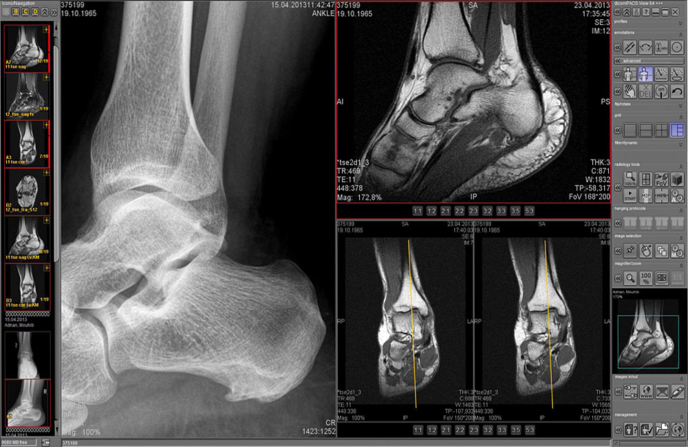
Digital images representing the pattern of incident X-rays – whether generated from direct or computed radiography systems – share a number of special characteristics. Digital signal detection and digital display offer new opportunities for regulating brightness and manually adjusting contrasts. As a result, details such as hairline fractures become apparent. Soft tissues can be visualised thanks to manual adjustments in the level of lightness and darkness within the same image.
Digitisation has a number of additional advantages. All images (X-rays, MRI, digital pictures) as well as documents (admission notes, findings reports, faxes, etc.) can be attached to patients’ medical records and are readily accessible. Backup and archive solutions guarantee quick access and data security in keeping with relevant medical products laws (depending on the manufacturer!).
Picture Archiving and Communication Systems (PACS) are user-friendly software solutions for administering the production and use of X-ray images: from exposure to image analysis, archival storage and file sharing.
The software enables users to quickly and easily share and view images. Depending on the manufacturer, systems can include numerous advanced features including electronic fax, document management, operative reports and X-ray histories. Software modules for 3D reconstruction, surgical planning, voice recognition, and statistics can also be integrated.
dicomPACS® by OR Technologies is a sophisticated, high-tech image management solution based on vendor neutral archive technology (VNA) and ideally suited for private practices and hospitals. With dicomPACS®, all images generated by digital X-ray, CT, MRI and ultrasound devices, as well as diverse documents (e.g., admission notes, medical findings, medical histories, faxes) are stored in a digital patient folder and readily accessible. Our carefully designed archive and backup solutions guarantee quick access and data security in keeping with the relevant medical devices act. Furthermore, the software can easily be integrated into all common information management systems (e.g., KIS, RIS and EPA). For more information, click here.
| Fast |
Immediate access to all digital patient records including X-ray images as well as documents from hospitals and doctor‘s offices |
| Cost-effective |
Digital radiography saves time and materials |
| No chemicals |
Some chemicals used in conventional radiography represent an occupational health hazard, are irritating and have an unpleasant odour |
| No loss of information |
Lost hard copies of X-ray images or patient records are a thing of the past |
| Space saving |
Archival storage of analog film material is costly in terms of space, money and time |
| Efficient communication |
Efficient communication between institutions and staff via various networks and the internet |
| Improved diagnostics |
Excellent images and a multitude of image processing options |
Software interface
PACS can interface with all administration software systems. This integration allows immediate access to patient data when submitting an X-ray order or recalling images from the archive. Interfaces can be established for GDT, BDT, HL7 and DICOM standards, as well as vender-specific systems.
Perfectly integrated
Imaging equipment for X-ray, ultrasound, MRI and arthroscopic examinations can be integrated into the existing computer network. Furthermore, faxes and other digital documents can be attached to the patient record. With the appropriate software solution, all information for a given patient is available at the click of a button and can be shared with other caregivers. The digital coordination of all data pertaining to a patient means that, as required per law, the full medical record can be accessed for many years in the future.
Requirements
Existing hardware can readily be integrated into the new system. Additional components such as printers, X-ray film scanners and other hardware can be purchased and integrated into the network at a time. Please consult your PACS provider concerning the specifications and requirements for servers, work stations and monitors.